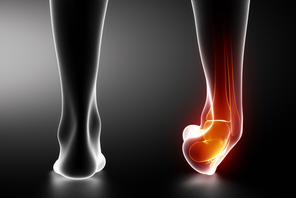
The most common ankle injuries involve lateral ligament damage and are one of the most prevalent seen by physiotherapists. Approximately 7-10% of emergency department hospital admissions are due to ankle strains1. The lateral ankle ligament is a complex of three different ligaments including the calcaneofibular ligament (CFL), anterior talofibular ligament (ATFL), and the posterior talofibular ligament (PTFL).
The most frequent inversion injury involves the CFL and ATFL. Normally the CFL prevents the ankle from rolling on its side inwards, while the ATFL stops the ankle from sliding forward. The medial ligament is a lot stronger than the lateral ligament and is consequently injured less frequently.
Contents
Causes of a torn lateral ankle ligament
Like most ligament injuries, the most common cause is sports related. Ankle sprains cause stretching or tearing of the ligament. Generally, a minor strain will only stretch the ligament. Tears may partially affect some of the ligament strands, or result in a complete rupture. The degree of the strain will determine how weakened the ligament is. Inversion injuries are the primary cause of torn lateral ankle ligaments as the ankle tilts inward, forcing all the pressure of the body weight onto the ankles’ outer edge. Consequently, the outside ligaments are stretched and potentially torn.
A ankle syndesmosis injury is a severe form of ankle sprain that also causes damage to other ligaments that support the ankle. Because this injury involves ligaments located above the ankle joint it is sometimes called a high ankle sprain. This injury affects at least one ligament that connects the fibula and tibia bones being sprained. Mild injuries of this type can take twice as long to heal compared with a typical ankle sprain.
Signs, symptoms and diagnosis of a torn lateral ankle ligament
A torn lateral ankle ligament presents as painful swelling and can often be ecchymotic. The bruising and swelling result from the rupturing of blood vessels caused by tearing of the soft tissues. Consequently, the initial swelling is due to bleeding into the surrounding tissues and the build-up of fluid.
Often people that suffer a sprained ankle will experience future strains due to ankle instability. This is especially the case for people that have received several minor sprains or a single severe sprain. When ligaments are sprained they can become thickened and irritated, further agitating the ankle.
Diagnosing torn lateral ankle ligaments is usually through physical examination and x-rays to identify any potential fractures. During the assessment the physician will determine:
- The degree of instability
- Loss of strength – Resisted eversion assessment
- Loss of range of motion (ROM): Dorsiflexion and Plantarflexion – Eversion and Inversion
- Level of reduced proprioception
If a very severe sprain is suspected, the physician may request an MRI to fully assess the extent of the injury and determine the recommended course of action.
The ankle injury will be classified based on the grades outlined below
Grade 1: Minimal swelling and tenderness due to microscopic collagen fibre tears. Impairment is minor. No casts or splits are needed and weight bearing can continue as tolerated.
Grade 2: Moderate tenderness and swelling, possible instability, and a decrease in the range of motion. Impairment is moderate, with only some complete tears of collagen fibres. Typical treatment may include immobilisation with a splint and physical therapy to strengthen and stretch the ankle.
Grade 3: Severe impairment associated with complete rupture/tear of the ligament. Significant tenderness and swelling. Immobilisation is necessary, along with physical therapy and possible surgery.
Treatment options for a torn lateral ankle ligament
The first response of any ligament sprain is to apply first aid immediately after the injury. This should involve Rest, Ice, Compression and Elevation (RICE). This helps to minimise bleeding at the site of the strained or torn ligament and can help to reduce discomfort and improve recovery time.
Rest: It’s important to avoid weight bearing and any other strenuous activity involving the ankle. Resting will help to minimise the potential for further damage.
Ice: Placing a cold pack on the site of the injury will reduce tissue metabolism2, and constrict the blood vessels to minimise swelling. Ten minute ice treatments have been shown to be most effective3.
Compression: To prevent further swelling (odema) from inflammation and bleeding a elasticated bandage should be applied to the ankle. This should be tight, but comfortable. Any bandaging should start distal to the injury, moving proximally by overlaying each prior layer by half.
Elevation: Raising the leg above the pelvis level serves to stop odema by lowering hydrostatic pressure and enhancing the venous return to the systemic circulation. This also helps to support the removal of waste products from the injury site.
There are variations of the RICE application; however the principles remain the same. The aim is to minimise pain and odema, plus improve recovery time and limit the risk of further injury.
Most sprains only require four to six weeks to heal, depending on the grade. If correctly immobilised, a compete ligament tear can heal without the need for surgery. In many instances, ankle stability can still be functional even with a torn lateral ligament because there are other overlapping tendons that provide motion and strength.
Grade 1 and Grade 2 sprains should effectively heal using the RICE approach. In the case of a Grade 3 sprain there may be some degree of permanent instability that requires a cast or brace for several weeks to allow the ankle to properly heal. Rarely is surgery required.
Ankle recovery typically follows three phases:
Phase 1: A week of resting to protect the ankle and limit swelling.
Phase 2: One to two weeks of gentle exercising to restore motion, flexibility and strength.
Phase 3: Several weeks to months of gradual return to normal activities, while avoiding twisting or turning the ankle.
Approximately 80% of acute ankle sprains will recover fully with conservative management, while the remaining 20% will develop into functional or mechanical instability4.
Torn lateral ankle ligament surgery
 Injuries that fail to respond to non-surgical treatments or present persistent instability may require surgery. There are two options:
Arthroscopy: An incision may be made to examine the joint and determine if any part of the ligament is caught within the joint or there are any loose bone fragments. While this is an exploratory procedure, if problems are identified they are usually repaired at the same time as the arthroscopy.
Reconstruction: The torn ligament is repaired with sutures or stitches, or other ligaments and/or tendons are used to repair the tear.
The aim of surgery is to tighten ligaments and ensure that they are properly attached to the bone. Severely damage ligaments are replaced. The most common procedure used is the Broström-Gould anatomic repair. This type of operation is often recommended following an arthroscopic joint inspection.
During this procedure a C or J-shaped incision is made over the ankle and ligaments identified. Using anchors or stitches attached to the fibula bone, these ligaments are tightened. Other tissue may be stitched over the ligaments to strengthen the repair. If the ligaments need replacing, tendons can be taken from the hamstring or a portion of the ankle and weaved into the bone surrounding the ankle, held in place with a screw or stitches.
This form of anatomic repair has been shown to improve ankle stability in high-demand athletes, especially when using suture anchors5. Li and colleagues suggest that this method can restore pre-injury functional level, although their research only assessed 62 patients.
Other research also published at the same time as Li and colleagues suggests that arthroscopic repair of chronic lateral ankle instability can be just as successful as a Broström-Gould anatomic repair. Nuro and Moreira used an anchor placed in the fibula during arthroscopic lateral ligament repair to treat 31 patients6. Although some complications were noted, most of these were minor and the authors reported overall success using this technique.
Rarely is surgery recommended for torn lateral ankle ligament injuries. Investigations by Prisk and colleagues suggests that neither repair or reconstruction will fully restore the contact mechanics of the joint, or the motion patterns of the foot7. Based on this research, Prisk and colleagues have identified a need for conducting long-term, prospective, comparative, in vivo studies to determine the appropriateness of operative treatment in comparison with- non-surgical responses.
Studies confirm that a prolonged history of instability of the lateral ankle ligaments can promote cartilage breakdown and subsequent osteoarthritis, although the severity is also influenced by ages and other risk factors8.
Prevention and recovery
Ankle sprain and associated ligament tears can be difficult to prevent, especially for individuals that play a lot of sport or are obese. The best way to protect the ankle is by maintaining good muscle flexibility, balance and strength. Wear appropriate supporting footwear, pay attention to working, running and walking surfaces, and warm-up prior to vigorous physical activities.
Nutrients supporting ligament recovery
During the recovery phase of any ligament injury it’s important to try take measures to promote healing and lessen inflammation. In addition to following the RICE guidelines, recovery can be aided by paying close attention to diet.
Good nutrition can help to accelerate healing by reducing tissue inflammation and stress. Eating plenty of fresh fruit and vegetables will help to increase antioxidant activity and lower inflammation.
Research by Barker and colleagues at the Sports Science Department of the Orthopedic Specialty Hospital in Utah found that people who took antioxidant supplements had a much higher rate of recovery after ligament damage compared with the control group9. This study confirmed that a higher intake of antioxidants assisted with the decrease in inflammatory agents, allowing the tissues to repair faster.
Vitamin C, protein, zinc and copper are all important nutrients that support ligament recovery. As vitamin C is involved in collagen production it is an essential precursor for the structural base of ligaments. Vitamin C has also been shown to successfully stop inflammation following surgery to repair torn ligaments10.
High-quality protein is also needed to facilitate rejuvenation of ligaments and other tissues following injury. Furthermore, zinc lowers oxidative stress and copper improves blood supply to the muscles, further supporting recovery11.
In addition to maintaining healthy diet, patients recovering from lateral ankle ligament injuries may also wish to take suitable supplements. There are a range of nutritional supplements that can help to ensure that the body has everything it needs to support healing.
References
- “van den Berkerom, M. et. al. (2012). Management of acute lateral ankle ligament injury in the athlete. Knee Surgery, Sports Traumatology, Arthroscopy. Volume 21, Issue 6, (pp. 1390-5).” ↩
- “Bleakley, C., McDonough, S., and MacAuley, D. (2004). The use of ice in the treatment of acute soft-tissue injury: a systematic review of randomized controlled trials. American Journal of Sports Medicine. Volume 32, Issue 1, (pp. 251-61).” ↩
- “Brucker, P. and Kahn, K. (2006). Clinical Sports Medicine, page 130.” ↩
- “Chan K. et. al. (2011). Acute and chronic lateral ankles instability in the athlete. Bulletin of the NYU Hospital for Joint Diseases. Volume 69, Issue 1, (pp. 1-26).” ↩
- “Li, X. et. al. (2009). Anatomical Reconstruction for Chronic Lateral Ankle Instability in the High-Demand Athlete: Functional Outcomes After the Modified Broström Repair Using Suture Anchors. American Journal of Sports Medicine. Volume 37, Issue 3, (pp. 488-94).” ↩
- “Nuro, C. And Moreira R. (2009). Arthroscopic repair of chronic lateral ankle instability. Foot and Ankle International. Volume 30, Issue 3, (pp. 213-17).” ↩
- “Prisk, V. et. al. (2010). Lateral Ligament Repair and Reconstruction Restore Neither Contact Mechanics of the Ankle Joint nor Motion Patterns of the Hindfoot. The Journal of Bone and Joint Surgery. Volume 92, Issue 14. (pp.2375-86).” ↩
- “Sugimoto, K. et. al. (2009). Chondral Injuries of the Ankle with Recurrent Lateral Instability: An Arthroscopic Study. The Journal of Bone and Joint Surgery. Volume 91, Issue 1, (pp. 99-106).” ↩
- “Barker, T. et. al. (2009). Antioxidant supplementation lowers circulating IGF-1 but not F(2)-isoprostanes immediately following anterior cruciate ligament surgery. Redox Report, Volume 14, Issue 5, (pp. 221-6).” ↩
- “Barker, T. et.al. (2009). Modulation of inflammation by vitamin E and C supplementation prior to anterior cruciate ligament surgery. Free Radical Biology & Medicine. Volume 45, Issue 5, (pp. 599-606).” ↩
- “Speich, M. et.al. (2001). Minerals, trace elements and related biological variables in athletes and during physical activity. Clin Chim Acta. Volume 312, Issue 1-3, (pp. 1-11).” ↩


