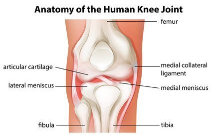
There are several ligaments that are vital for the stability of the knee joint. One of these is the medial collateral ligament (MCL).
This ligament is located on the inner edge of the knee and helps to secure the inside of the joint and stop the knee from ‘opening up’. It joins the inner aspect of the shin bone (tibia) with the inner aspect of the thigh bone (femur). This ligament provides stability by preventing valgus forces and excessive twisting.
This short video shows the motion of the MCL in flexion-extension movement.
Contents
Causes of a Torn MCL
A torn MCL is usually the result of an abnormal twisting movement when combined with excessive pressure, such as landing awkwardly from a jump. It’s also possible to tear the MCL by impacting the outer knee and forcing it to bend in the opposite direction. While it is most commonly associated with a specific event, repetitive strain may also cause the MCL to tear.
MCL injuries often are inflicted from sports that require fast changes in direction or contact. These may include netball, football, skiing, and basketball. Repetitive activities like kicking during breaststroke swimming may also damage the MCL.
Signs, Symptoms and Diagnosis of a Torn MCL
There are several symptoms and signs of a torn MCL:
- At the time of the injury there is an audible tearing or snapping sound
- Pain is concentrated to the inner part of the knee
- There is stiffness and swelling of the knee
- The knee joint feels loose
- There is knee instability and it won’t bear weight
A torn MCL may range in severity from a small partial tear with minimal pain, through to total rupture causing significant pain and immobility. The following grading is often applied to characterise the severity of a MCL tear:
Grade 1: Full function remains, although a few fibres are torn and there is minor discomfort.
Grade 2: There is moderate loss of function as a result of a significant number of torn fibres.
Grade 3: Knee instability occurs as all fibres are ruptured causing major loss of function. This injury may also damage other structures such as the cruciate or menisci ligaments.
A thorough examination from a physiotherapist is often all that’s needed to confirm a MCL. In some cases a X-ray, MRI or CT scan may be necessary to confirm the diagnosis and the extent of injury.
With correct management, grade 1 and 2 MCL injuries will heal within 2-8 weeks. Longer rehabilitation periods are necessary for a complete MCL rupture, especially if other structures of the knee are also damaged.
Treatment Options for a Torn MCL
There are a range of treatment options for torn MCL:
- Joint manipulation and/or mobilisation
- Soft tissue massage
- Bracing
- Taping
- Heat or ice treatment
- Anti-inflammatories
- Electrotherapy
- Hydrotherapy
- Biomedical correction
- Hydrotherapy
- Specific exercises to enhance strength, balance and flexibility
- Education
There are several products that can be used to help in the recovery of a torn MCL. Ice or hot packs, self massage foam rollers, protective/supportive tape, and resistance bands are some of the beneficial products.
MCL Surgery
 Until recently, knee medial collateral ligament surgery was largely focused on autograft techniques. This is a surgical reconstruction procedure that utilises the patient’s own tissue. It has been the preferred method because fast-healing tissue can be used to accelerate the recovery time. However, there are risks linked to this process because a second surgical site is required for tissue harvesting and this can present other complications.
A recent publication suggests that the use of an Achilles allograph combined with anatomic insertions is a preferred surgical treatment. Marx evaluated 14 patients that had undergone isometic reconstruction of the MCL, assessing knee ligament laxity, motion, functional outcome scores, and activity level scores. The time period for the follow-up was between 24 and 61 months1. This technique was shown to be very successful and enabled patients to return to pre-injury activity level.
New research has compared the effectiveness of anatomic MCL augmented versus anatomic reconstruction. Prior to this study, there has been no biomedical evaluation or comparison between these treatments for grade 3 injuries. Wijdicks and colleagues concluded that both treatments provided equivalent joint stability and enhanced knee kinematics2.
New animal based research indicates the potential for platelet-rich plasma to enhance tissue healing following an MCL injury. This Japanese-based study assesses rabbit knees with MCL tears. These joints were either left untreated or received a local delivery of plasma rich in growth factors (PRGF-Endoret). The findings provided evidence that PRGF-Endoret enhances the early stages of tissue healing3.
However, the treatment protocols and manufacturing procedures associated with platelet-rich plasma make assessing the tissue healing efficacy difficult. Further research is required.
Prevention and recovery
Education: Knowing how to correctly land or fall can assist in the prevention of MCL injuries. For example, falling sideways or forward rather than backwards can help to reduce the stress on knee joints. People participating in sports such as skiing, basketball, football and soccer should practice landing/falling correctly as part of their training.
Stretching: Muscles and ligaments should be prepared for aerobic activity by engaging in proper warm-ups. This helps to minimise any shock from sudden motion. Three to five minutes of light aerobic exercise is usually sufficient. Stretching the muscles around the knee after exercise is also important for preventing tightening of the MCL during recovery.
Strengthening: Maintaining strong muscles around the ligaments and knee joint can help to reduce the strain on the MCL. Strong quadriceps muscles are particularly beneficial. Located above the knee, these muscles are used in running and can be strengthened with squats and lunges.
Rest: The MCL can wear out and be stretched over time. Thus, it’s important to allow the body time to recover. Human knee joint anatomy. Alternating between high and low-impact activities can help to minimize the strain on the knee.
Nutrients Supporting Ligament Recovery
Nutrition plays a big role in the recovery following a serious injury to ligaments. Certain vitamins and minerals can help to speed up the process. Vitamin C, zinc, copper, and omega-3 fatty acids are some of the key nutrients.
Boosting antioxidant activity helps to reduce inflammation and supports healing processes by limiting oxidative stress. Supplements and a diet enriched with high-quality protein, and fresh fruit and vegetables can be very beneficial.
References
- “Marx, R. (2012). Surgical technique: medical collateral liaament reconstruction using Achilles allograft for combined knee ligament injury. Clinical Orthopaedics and Related Research. Volume 470, Issue 3, (pp. 798-805).” ↩
- “Wijdicks, C. et.al. (2013). Superficial medical collateral ligament anatomic augmented repair versus anatomic reconstruction. An In Virtro Biomechanical Analysis. American Journal of Sports Medicine. Volume 41, Issue 12, (pp. 2858-66).” ↩
- “Yoshioka, T. et.al. (2012). The effects of plasma rich in growth factors (PRGF-Endoret) on healing of medical collateral ligament of the knee. Knee Surgery, Sports Traumatology, Arthroscopy. Volume 21, Issue 8, (pp. 1763-69).” ↩


